Comprehensive Eye Exam
Our Doctors will use a wide variety of tests and procedures to examine your eyes. The purpose of these tests is to determine not only your ability to see, but also to evaluate the overall health of your eyes. Many of the tests performed during an eye exam are similar, but testing will vary depending on your individual needs.
- Your exam may include automated testing to give the doctor baseline information about your eyes. This includes autorefraction – a test to determine if you are nearsighted, farsighted or have astigmatism, lensometry – a test to read the current prescription in your glasses, and keratometry – a test to determine the curvature of your cornea.
- Your vision will be measured by reading the eye chart or visual acuity.
- Muscle function tests will be performed to check the movement of your eyes. This test will identify any muscle weakness or involuntary eye movement. We will check your binocular vision to ensure your eyes work together. Binocular vision or stereo vision is important for proper depth perception, eye muscle coordination and the ability to change focus from near to far objects.
- Visual field testing may be performed to measure your peripheral vision, or the total area you are able to see. This test is used to detect and monitor diseases of the eyes and neurological disorders.
- A check of your eye pressure or intraocular pressure will be performed. High intra-ocular pressure may be a sign of glaucoma.
- Color vision testing will allow us to ensure you perceive colors properly.
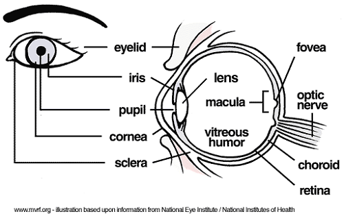
- A total eye health assessment will be performed. This will allow the doctor to evaluate the overall health of your eyes. We will evaluate your pupil responses, cornea, lens, vitreous, retina, optic nerve, macula and more.
Typically an eye exam will take anywhere from a half hour to an hour depending on the number and complexity of tests required for your eyes.
EYE ANATOMY